Chapter/Index: Introduction | A | B | C | D | E | F | G | H | I | J | K | L | M | N | O | P | Q | R | S | T | U | V | W | X | Y | Z | Appendix
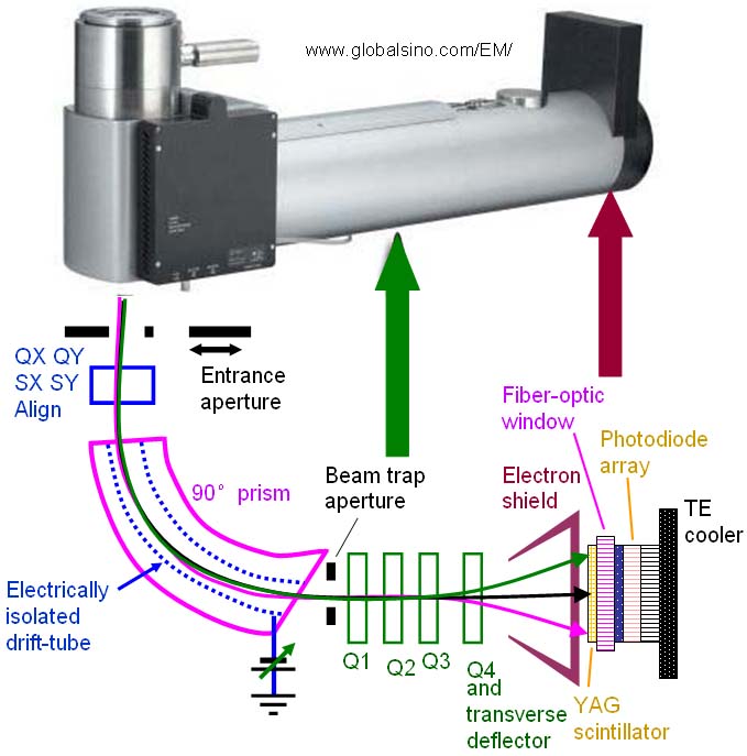
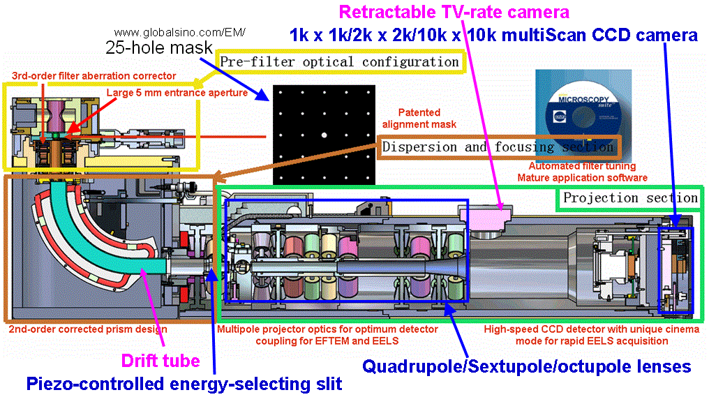
| The development of parallel EELS detectors, specifically multichannel arrays, has allowed a large gain in the collection efficiency, typically by a factor of 500, compared with the earlier serial spectrometers. A parallel EELS spectrometer (PEELS) collects the whole energy spectrum simultaneously since it does not use slits or maximally widens the slit, and thus it is more efficient than SEELS due to its higher rate of data acquisition. A PEELS uses a YAG scintillator and a position–sensitive photon detector such as a linear, parallel photodiode array (or more commonly now using a scintillator-CCD combination), and thus the spectrum is recorded in parallel. In this way, the scintillator is coupled to the semiconductor array through fiber optics in the dispersion plane of the spectrometer, indicated in the schematic illustration of general Gatan PEEL spectrometers in Figure 4883a. On the other hand, a set of postfield lenses is typically used to magnify the energy dispersion on the dispersion plane before the electrons reach the scintillator, in order to allow the detector channels to properly read the spectrum. For this reason, a series of quadrupoles (normally four) are used to control both the dispersion and the width of the spectrum at the detector. The whole energy spectrum is read out via an amplifier through an A/D (analog/digital) converter and into a multi-channel analyzer system. The PEEL spectra with sufficient intensity can be acquired in < 1 second. Figure 4618b shows an advanced GIF Tridiem [1].
Figure 4883a. Top image: example of Gatan PEEL spectrometers; Bottom image: schematic of spectrometer. As an example, Figure 4883b shows an advanced GIF Tridiem. The parallel detection is obtained because the CCD has a diode array with 1024 or more diodes to collect the whole spectrum in parallel.
Figure 4883b. Schematic of GIF (Gatan Imaging Filter) Tridiem camera. Note that the post-column Gatan Image Filter (GIF) is a development of Gatan magnetic-prism PEELS.
|