
Chapter/Index: Introduction | A | B | C | D | E | F | G | H | I | J | K | L | M | N | O | P | Q | R | S | T | U | V | W | X | Y | Z | Appendix
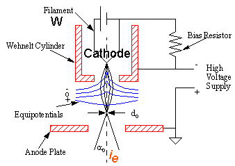
| The current density of electron gun is the number of electrons (or charge) per unit area per unit time. Although current density is an useful term, it is more important to define the brightness because brightness is the current density per unit solid angle of the source. Electron sources differ considerably in their size and, as a result, the electrons leave the source with a range of divergent angles, so we can’t ignore the angular distribution of the electrons. In analytical electron microscopy (AEM) and STEM, brightness is particularly important when we are using very small convergent probes. The concept of brightness is less important in conventional TEM where we use a relatively large, defocused beam, but it is still relevant to the intensity we see on the screen, and so it affects how easy it is to operate the microscope and see our images and DPs. The characteristics of electron source, defined at the gun
crossover, that is, the point at which the electrons are
focused after leaving the source (as shown in Figure 4961 (a) below):
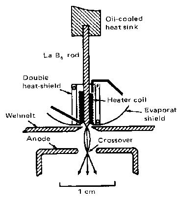
The current density (current per unit area) is ie/π(d0/2)2 and the solid angle of the source is πα02, so the brightness β (units: A/m2 sr.) is The β is an invariant quantity in any electron-optical system by changing the solid angle of the source. It is important that the higher the value of β, the more electrons we can put into a beam of a given size, and so the more information we can generate from the specimen. On the other hand, the higher the value of β, the more we will damage sensitive specimens. Actually, the β can be directly given by the brightness of the emitting cathode (βC) and the accelerating voltage VA: This ideal value β in Equation 4961b can only be reachable if the cathode tip is well aligned with the aperture of the Wehnelt cylinder and a proper bias voltage is applied to the cylinder. A table on the comparison of various electron sources, including their brightnesses, is presented in page1409. Furthermore, with high-brightness electron sources and aberration correctors, very large current density (> 106 A/cm2) in electron microscopes (EMs), especially in TEMs, can be obtained within a small electron probe in diameter (< 1 nm). In EMs, the condenser aperture is used to exclude electrons emitted at high angles from the electron gun, which will decrease the brightness but improve the quality of the illumination because these peripheral electrons are less coherent, especially in LaB6 and W (tungsten) guns. The most promising coherent electron sources for electron microscopes (EMs) have been found in ultrasharp nanotips [1–3] in field emission electron guns due to the very high coherence and significant brightness. Table 4961. Function of "BRIGHTNESS" knob in TEM systems .
|
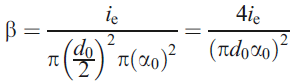 --------------------------- [4961a]
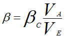 --------------------------- [4961b]