Chapter/Index: Introduction | A | B | C | D | E | F | G | H | I | J | K | L | M | N | O | P | Q | R | S | T | U | V | W | X | Y | Z | Appendix
| Electron energy loss spectroscopy (EELS) measures the spectral distribution of energy transferred from an incident electron beam into a specimen. In general, there are mainly two types of fundamental methods: EELS is related to electronic, optical and mechanical properties of the observed materials. Methods i) and ii) above are also called reflection electron energy loss spectroscopy (REELS) and transmission electron energy loss spectroscopy (TEELS) modes, respectively. However, different from REELS, the name of TEELS is conventionally simplified as EELS. REEL spectrum is acquired with a reflected electron beam by the EELS spectrometer. More than 50% of the reflected electrons experience multiple valence losses. The REEL spectrum displays the intensity distribution of the reflected electrons as a function of the energy loss and is particularly sensitive to the electronic structure of surfaces. REELS presents several advantages with respect to TEELS, for instance, the use of incident electron beam energies lower than 3 keV enable the measurements performed in conventional ultra-high-vacuum systems normally used for photoemission and Auger spectroscopy [1] and there is no constraint of sample thickness [2]. In REELS (reflection electron energy loss spectroscopy) measurements, the electrons follow many different trajectories inside the solid within its surface region, and all of them contribute to the cross section of inelastic electron scattering. Reflection energy-loss spectroscopy (REELS) can be performed with 30-keV electrons in a molecular beam epitaxy (MBE) chamber for surface analysis during crystal growth. REELS is also possible in a TEM at 100 keV or more. Figure 2302a shows the REELS spectra for the Al2O3/SiO2 thin films on Si substrates, before and after annealing, at a primary energy of 1.0 keV. The onset due to electron–hole excitation corresponds to the band gap values of the dielectric films. Therefore, the band gap energy is given by the crossing point found by drawing a linear fit line from a point near the onset of the loss spectrum to the background level. [3 -5].
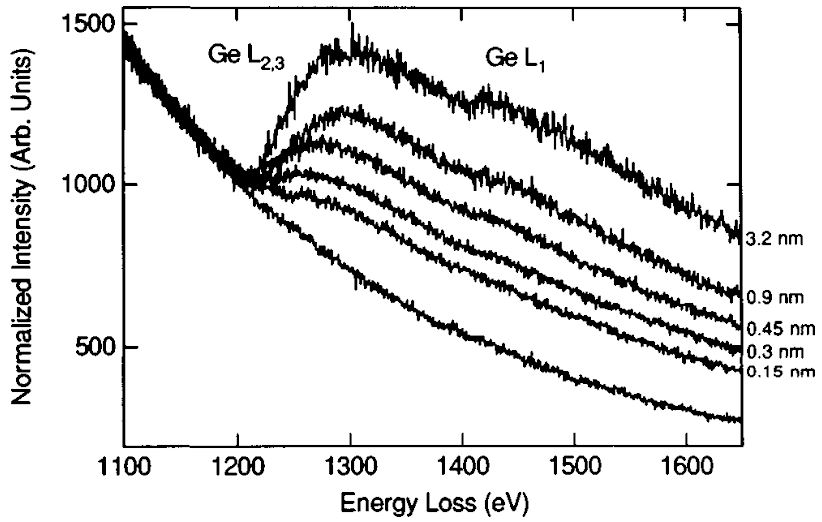
Figure 2302b shows REELS spectra recorded during in situ MBE growth of Ge on Si, illustrating the surface sensitivity of REELS, that is, the change of the intensity of Ge L2,3 edge with the increase in thickness of the deposited Ge layer.
[1] J.C. Ingram, K.W. Nebesny, J.E. Pemberton, Appl. Surf. Sci. 44
(1990) 279.
|