| In order to interpret the Kikuchi bands on a given EBSD pattern in terms of atomic geometry in the crystal, a reference frame for the image is required. The position of pattern center (PC) of EBSD is described by the point of intersection of a sphere of reflection with the plane of the photograph and indicates the relative position of the image with reference to the interaction volume of the sample.
The pattern center (PC) in EBSD is critical for accurately interpreting the Kikuchi bands and understanding the atomic geometry of the crystal. The techniques that can be used to locate PC on an EBSD are:
- Conic fitting or three steel-ball method:
- This method involves placing three steel balls in the EBSD sample chamber. Their known positions relative to the pattern allow the construction of a conic section that fits the Kikuchi pattern. By comparing this known geometry with the captured pattern, the PC can be accurately determined.
- Circular mask method:
- A mask with a circular pattern is used to project onto the EBSD detector. By matching the geometry of the known circular mask to features in the pattern, the PC can be located. This is a relatively simple method that uses known geometrical shapes to determine the center.
- Known orientation method:
- In this technique, a crystal with a known orientation is used, and the expected Kikuchi band pattern is compared to the observed one. By aligning the predicted bands with the actual bands, the PC can be adjusted and located.
- Shadow casting [1]:
- This method uses a shadow projected by the electron beam interacting with the sample surface. The shadow’s position relative to the Kikuchi bands gives clues about the PC’s location. Adjusting the sample or beam orientation helps in finding the correct PC position.
- Iterative fitting [2]:
- This is an optimization method in which an initial guess of the PC is adjusted iteratively. By comparing the theoretical Kikuchi pattern to the experimental one, the PC is fine-tuned through multiple iterations until the best fit is achieved.
- Screen moving [3]
:
- In this method, the EBSD screen (detector) is physically moved to different positions, and the shifts in the Kikuchi patterns are observed. By analyzing how the pattern shifts with screen movement, the location of the PC can be deduced.
Each of these methods serves to establish an accurate reference frame for the EBSD pattern, ensuring that the Kikuchi bands can be interpreted correctly in relation to the crystal structure of the sample.
These techniques offer accuracy ranging between 1% to 0.2% of the phosphor width for different PC components. Figure 2338a shows the pattern center for band identification in EBSD analysis.Here, the angle θ, prescribing the band width, follows the Bragg's law λ = 2dhklsin θ, and dhkl represents the d-spacing for reflecting plane. z* is the distance from the specimen to the screen.
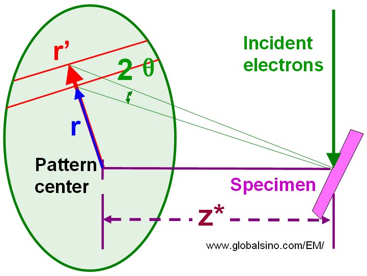
Figure 2338a. The pattern center for band identification in EBSD analysis.
Figure 2338b shows the formation of a Ge (germanium) EBSD pattern projected onto a sphere centered at the PC. These kinematically simulated Kikuchi bands (the gray lines enclosing the bright bands) had been widened by a factor of 2 from their Bragg angles to ensure that the EBSD bands are fully enclosed.
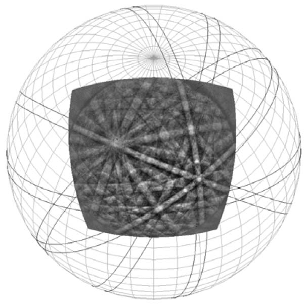
Figure 2338b. The formation of a Ge EBSD pattern projected onto a sphere centered at the PC. [4]
[1] Venables, J.A. & Bin Jaya, R. (1977). Accurate microcrystallography
using electron back-scattering patterns. Phil Mag 35(5),
1317–1332.
[2] Krieger Lassen, N.C. (1999). Source point calibration from an arbitrary electron backscattering pattern.J Microsc 195, 204–211.
[3] Carpenter, D.A., Pugh, J.L., Richardson, G.D. & Mooney, L.R.
(2007). Determination of pattern centre in EBSD using the
moving-screen technique. J Microsc 227 (September), 246–247.
[4] Jay Basinger, David Fullwood, Josh Kacher, and Brent Adams, Pattern Center Determination in Electron Backscatter Diffraction Microscopy, Microsc. Microanal. 17, 330–340, 2011.
|
