Intensity Distribution of HRTEM Image
- Practical Electron Microscopy and Database -
- An Online Book -

https://www.globalsino.com/EM/  


This book (Practical Electron Microscopy and Database) is a reference for TEM and SEM students, operators, engineers, technicians, managers, and researchers. You can click How to Cite This Book to cite this book. Please let Dr. Liao know once you have cited this book so that the brief information of your publication can appear on the “Times Cited” page. This appearance can also help advertise your publication.


=================================================================================

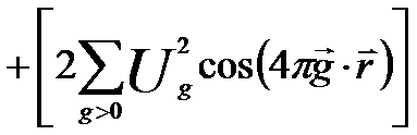
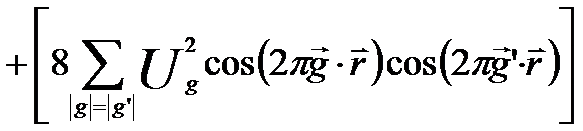
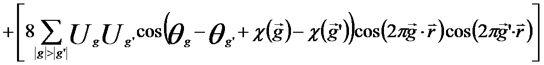
The local intensity distribution I(r) of HRTEM image is a function of the amplitudes and phases of the transmitted and diffracted electron beams of the exist wave from the TEM specimen. I(r) can be given by [1 - 2],

          local intensity distribution I(r) of HRTEM image ---------------------- [4145a]
                   local intensity distribution I(r) of HRTEM image
                    local intensity distribution I(r) of HRTEM image
                    local intensity distribution I(r) of HRTEM image
                    local intensity distribution I(r) of HRTEM image
                    local intensity distribution I(r) of HRTEM image ------------- [4145b]

where,
         ψi -- The complex electron wave on detector or photographic film
         U0 -- The amplitude of the transmitted electron beam
         Ug -- The amplitude of the diffracted electron beam
        θ0 -- The phase of the transmitted electron beam
        θg -- The phase of the diffracted electron beam
        χ(g) -- The phase change χ(g)

The phase change χ(g) reflects the influence of the contrast transfer function of the objective lens. The first term in Equation 4145b represents the background intensity of the HRTEM image while the other terms give cosine fringes of various periodicities and amplitudes due to the local intensity modulation produced by constructively and destructively interfere at the image plane.


[1] K. Ishizuka, Ultramicroscopy 5 (1980) 55.
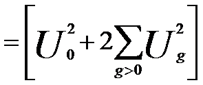
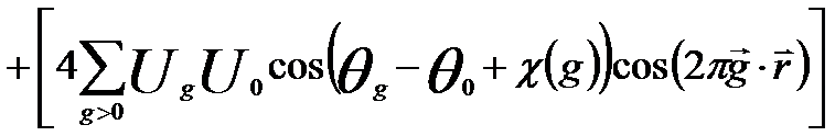
[2] R. W. Glaisher and A. E. C. Spargo, Ultramicroscopy 18 (1985) 323.


=================================================================================

The book author (Dr. Liao) welcomes your comments, suggestions, and corrections, please click here for submission.


Copyright (C) 2006 GlobalSino, All Rights Reserved