Chapter/Index: Introduction | A | B | C | D | E | F | G | H | I | J | K | L | M | N | O | P | Q | R | S | T | U | V | W | X | Y | Z | Appendix
|
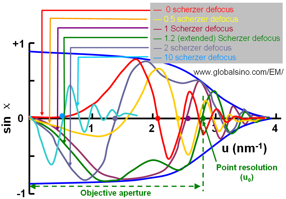
HRTEM usually operates under phase-contrast conditions because the information about the atomic structure of the object consists in the phases of the elastically scattered electron waves. In order to convert a small phase shift into a proper change of contrast, an extra phase shift must be added so that the phase difference between the scattered and unscattered wave is equal to 180°. In conventional TEMs this phase shift is introduced by defocusing the objective lens properly [1]. For HRTEM imaging, CTF (contrast transfer function) partially changes the contrast of the images by adjusting Scherzer defocus. Assuming Cs and λ are constant for a specific microscope, Figure 4232 shows CFT T(|g|) = sin(χ) at n Scherzer defoci, given by, where, n -- 0, 0.5, 1, 1.2 (extended), 2, and 10
Figure 4232. Contrast transfer function (CFT) at 0, 0.5, 1, 1.2 (extended), 2, and 10 Scherzer defoci. Here, 1.2 scherzer defocus is extended Scherzer defocus, which normally is the best defocus condition to take HRTEM images (see page2779). In this case, Cs and Δf are balanced and all diffracted beams have nearly constant phase out to the first zero. Indicated by the green curve in Figure 4232, to maximize the transferred information about the TEM sample in a single image it is necessary to set the defocus C1 (Δf) to an optimum negative number (negative 1.2 Scherzer defocus) to compensate for the effects of the spherical aberration, C3 (Cs) and thus, have a largest interval from 0 to a maximum spatial frequency, u0, where sin(χ) ≈ -1. Figure 4232 also shows that the aperture function (defined by the objective aperture) has been set to cut off the CTF after the first crossing of the u-axis where it starts to oscillate rapidly at this defocus. Although the information greater than u0 is not included, it makes more easily to interpret the images since they do not include the information out of phase. The spatial frequency, u0, where the phase contrast transfer function crosses the u-axis the first time at the optimum defocus setting defines the point to point resolution of the microscope.
[1] O. Scherzer, J. Appl. Phys. 20 (1949) 20.
|