Chapter/Index: Introduction | A | B | C | D | E | F | G | H | I | J | K | L | M | N | O | P | Q | R | S | T | U | V | W | X | Y | Z | Appendix
|
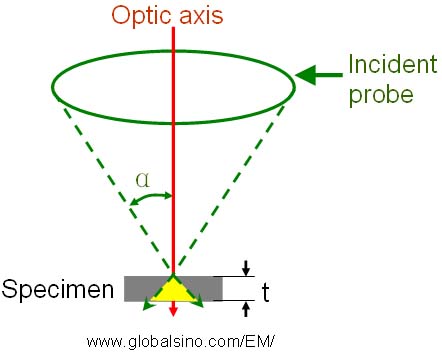
Combined magnetic and electrostatic quadrupoles can create negative chromatic aberration and octupoles generate aperture aberrations in correcting both spherical and chromatic aberrations for a resolution limit of 1 nm at low accelerating voltages between 0.5 and 1 keV for SEM [1]. Most modern scanning electron microscopes (SEMs) can record surface morphology but does not provide atomic resolution. The first atomic resolution, Hitachi aberration-corrected electron microscope, HD-2700C (page4444) was installed at Brookhaven National Laboratory [2]. Furthermore, Figure 4244 shows the schematic illustration of interaction volume with a thin film as the SEM specimen. It is clear that the interaction volume is larger when the semi-convergence angle of the SEM probe is larger, and thus the SEM spatial resolution will be affected. Therefore, in order to optimize the spatial resolution, it is important to balance the following factors:
Figure 4244. Schematic illustration of interaction volume (in yellow) with a thin film as a SEM specimen. α is the semi-convergence angle, while t is the specimen thickness.
[1] Zach, J. and Haider, M., 1995. Aberration correction in a low voltage SEM by a multipole corrector, Nucl. Instrum. Methods Phys. Res., A363, 316–325. [2] Y. Zhu, H. Inada, K. Nakamura, and J. Wall, Imaging single atoms using secondary electrons with an aberration-corrected electron microscope, Nature Materials, 8 (2009) 808.
|