Chapter/Index: Introduction | A | B | C | D | E | F | G | H | I | J | K | L | M | N | O | P | Q | R | S | T | U | V | W | X | Y | Z | Appendix
| Similar to optical prism as shown in Figure 4992a, the basic electron spectrometer (e.g. GIF, EELS, and PEELS detectors) is the prism spectrometer as shown in Figure 4992b. In this curved prism, electrons are deflected by about 90° in a perpendicular uniform static magnetic field B (of the order of 0.01 T). According to the Lorentz force law, the deflection angle depends on the electron velocity and strength of magnetic field. Therefore, in the curved magnetic field, electrons with different energies are dispersed, and consequently an EEL spectrum is obtained at the end of this part. In fact, the magnetic prism is the same as in a parallel electron energy loss spectrometer, and, of course, an EEL spectrum can be measured with the GIF as well. The detector in the spectrometer can be an array of photodiodes or a charge-couple device (CCD), in both cases combined with a suitable scintillator and a light-guide (e.g. fiber).
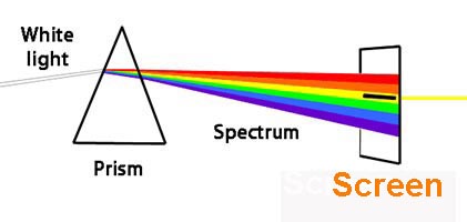
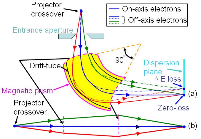
Figure 4992a. Schematic showing optical prism. In Figure 4992b (a), the spectrum is formed in the dispersion plane, consisting of a distribution of electron counts (I) versus energy loss (ΔE). All the electrons suffering the same energy loss but traveling in both on-axis and off-axis directions are directed to a focus in the dispersion plane of the spectrometer, which acts as a homogenous magnetic lens as shown in the equivalent schematics in Figure 4992b (b). The object plane of the spectrometer is typically set at the back focal plane of the projector lens.
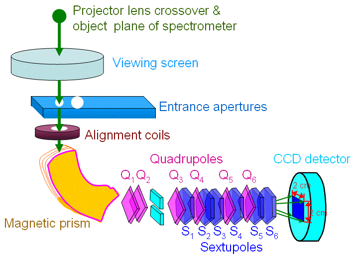
Figure 4992b. (a) Schematic showing magnetic prism, and (b) Equivalent schematics of the magnetic prism. Electrons at various kinetic energies (due to energy losses induced by interaction with TEM specimen) are focused at the energy-dispersive plane of the spectrometer. For the most common EFTEM unit as shown in Figure 4992c, the detection system consists of a slow scan CCD array detector rather than a single line of diodes used in PEELS detector.
Figure 4992c. Gatan imaging filter using CCD detector.
By following Equation [4919c], the electrons follow circular paths of radius R is given by R = (m/e)Bv -------------------------------------------- [4992] A limitation of the magnetic prism-based EELS detection system is the
spectrum drift in the energy dispersion direction. Various sources of instability
|