Chapter/Index: Introduction | A | B | C | D | E | F | G | H | I | J | K | L | M | N | O | P | Q | R | S | T | U | V | W | X | Y | Z | Appendix
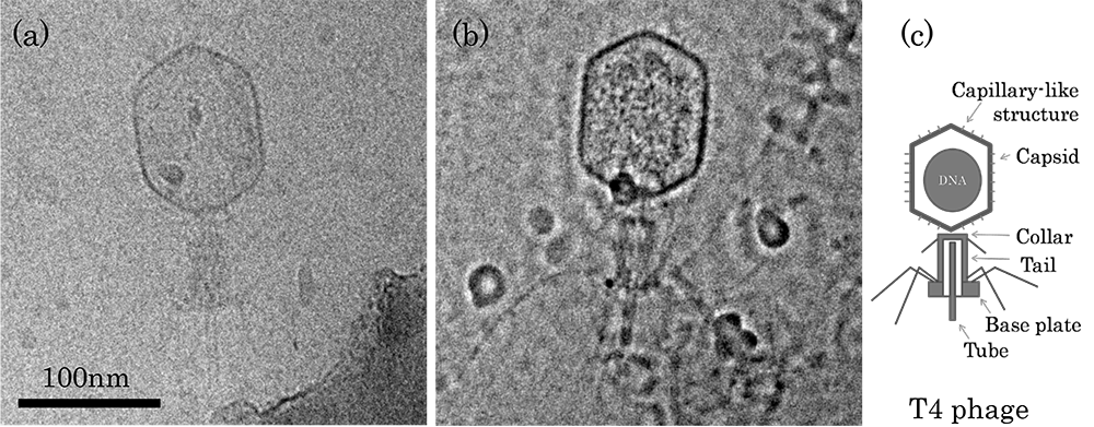
| In many cases, the existence of interfacial layers cannot be shown in conventional bright-field TEM images because they all are in the amorphous phase and have similar atomic numbers. In this case, defocus function can be used, which is called Zernike phase contrast. In TEM technique, Zernike phase contrast can be obtained by converting the phase change of electron waves, scattered by a specimen, into the amplitude change. The conversion can be carried out by either using a Zernike phase plate or a combined effect of the defocus and spherical aberration of the electron lens. In the later method in TEM operation, one only needs to simply change the defocus since any microscope has spherical aberration (refere to Phase Shift from Defocus and Spherical Aberration). Figure 1203 shows TEM observation of an ice-embedded T4 phage. One can clearly see the fine structures of DNA in a capsid, the hair-like structural objects and the cylindrical structures on the capsid surface the image of Zernike phase contrast in Figure 1203 (b).
[1] www.jeol.co.jp.
|