| Determining whether a dislocation is a screw dislocation, an edge dislocation, or a mixed type involves analyzing the relationship between the dislocation line and its Burgers vector, as well as using specific techniques like transmission electron microscopy (TEM). Here are the steps and methods to distinguish between screw and edge dislocations:
- Burgers Vector Analysis:
- Edge Dislocation: The Burgers vector is perpendicular to the dislocation line. The dislocation is characterized by an extra half-plane of atoms that ends within the crystal, causing a distortion in the lattice.
- Screw Dislocation: The Burgers vector is parallel to the dislocation line. In this case, the crystal planes are twisted around the dislocation line, creating a spiral structure as the name "screw" suggests.
- TEM Imaging and Diffraction Contrast:
- Transmission Electron Microscopy (TEM): TEM is a powerful technique for directly observing dislocations in a crystal. The method relies on diffraction contrast, where dislocations appear as dark lines or regions in the image due to the strain they cause in the crystal lattice.
- Two-Beam Condition: By tilting the specimen in the TEM to a specific orientation (two-beam condition), the diffraction conditions can be adjusted to make certain dislocations visible or invisible, depending on their type.
- Screw Dislocation: In specific diffraction conditions, screw dislocations can appear with contrast in certain directions that edge dislocations would not.
- Edge Dislocation: Edge dislocations show contrast based on their interaction with the incident electron beam and the resulting strain field, which differs from that of screw dislocations.
- g·b Analysis (Visibility Criterion):
- The g·b criterion is used in TEM to determine the visibility of dislocations. Here, g is the diffraction vector and b is the Burgers vector. This criterion is used in TEM to determine whether a dislocation will be visible in a particular diffraction condition. If the g·b (where g is the diffraction vector and b is the Burgers vector) is non-zero, the dislocation will generally be visible.
- Edge Dislocation: For an edge dislocation, the dislocation will be visible in TEM if the dot product g·b is non-zero. The visibility is mainly influenced by the strain fields perpendicular to the dislocation line.
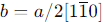
- For instance, to determine whether the threading edge dislocation with a Burgers vector of
a/2[1-10] can be visible under specific diffraction conditions, we'll use the g·b criterion. This criterion states that a dislocation is visible in a TEM image when the dot product of the diffraction vector
g and the Burgers vector
b is non-zero (g·b
≠
0).
- Calculate g·b for each of the given g vectors:
- For g = (200) case:


Therefore, for g = (200) case, g·b
≠
0, so the dislocation will be visible.
- For g = (101) case:


Therefore, for g = (101) case, g·b
≠
0, so the dislocation will be visible.
- For g = (002) case:


Therefore, for g = (002) case, g·b
=
0, so the dislocation will not be visible.
- Dislocation Line Observation:
- Screw Dislocation: The dislocation line appears as a continuous line when observed in a TEM micrograph under specific conditions.
- Edge Dislocation: The dislocation line often appears as a series of "steps" due to the presence of the extra half-plane of atoms, visible when the dislocation is viewed edge-on.
The Two-Beam Condition is a powerful and commonly used technique in transmission electron microscopy (TEM) to identify and characterize dislocations, but it is not the only method available. Several other techniques and approaches can also be used to distinguish between screw dislocations and edge dislocations: - Weak-Beam Dark-Field (WBDF) Microscopy:
- WBDF is a TEM technique that enhances the contrast of dislocations by imaging them using diffracted beams that are slightly off the exact Bragg condition (weak beams). This technique allows for more detailed observation of the dislocation core structure and is especially useful for visualizing the fine details of both screw and edge dislocations.
- High-Resolution TEM (HRTEM):
- HRTEM provides direct imaging of atomic columns and can be used to visualize the structure of dislocations at the atomic level. In HRTEM images, the difference in atomic arrangement around screw and edge dislocations can be observed, allowing for their identification:
- Edge Dislocation: Appears as an extra half-plane of atoms.
- Screw Dislocation: Appears as a helical or twisted arrangement of atoms.
- Dislocation Core Structure Analysis:
- By examining the core structure of a dislocation, which is the region near the dislocation line where the lattice distortion is greatest, it’s possible to differentiate between screw and edge dislocations. The arrangement of atoms around the dislocation core differs between screw and edge types, which can be observed in detailed TEM or HRTEM images.
- Selective Etching:
- In some materials, dislocations can be revealed by selective etching techniques where etchants preferentially attack regions of high strain. Edge and screw dislocations might etch differently, producing distinguishable surface features that can be observed under an optical microscope or a scanning electron microscope (SEM).
- Burgers Circuit:
- The Burgers circuit method involves tracing a closed loop around the dislocation in a TEM image and comparing it to a perfect lattice. The closure failure (the gap that appears) gives the Burgers vector directly. By knowing the direction of the dislocation line and the Burgers vector, the dislocation can be classified as edge, screw, or mixed.
- Edge Dislocation: The closure failure occurs perpendicular to the dislocation line.
- Screw Dislocation: The closure failure occurs parallel to the dislocation line.
- X-ray Topography:
- X-ray topography is a non-destructive imaging technique that can reveal dislocations in crystalline materials. Different types of dislocations (edge, screw) will produce different contrast features in the X-ray topographs due to their different strain fields.
|
