Chapter/Index: Introduction | A | B | C | D | E | F | G | H | I | J | K | L | M | N | O | P | Q | R | S | T | U | V | W | X | Y | Z | Appendix
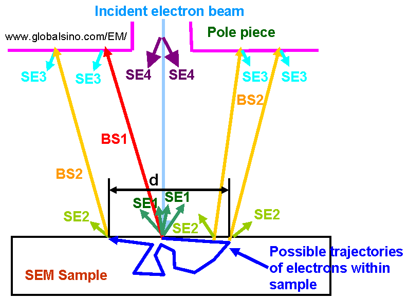
| There are four sources of secondary electrons (SEs): SE1, SE2, SE3, and SE4 as shown in Figure 4870. These electrons are classed according to how they are generated. A SE1 is electrons that are generated at the point of primary electron beam (PEB) impingement in surface of the specimen. Therefore, it carries the highest resolution information. A SE2 is electrons that are generated when a backscattered electron leaves the surface of the specimen. Due to the energy of backscattered electrons, this SE2 could leave the surface of the specimen sub-microns away from the PEB impingement site. SE2 add greatly to overall image brightness and generate sub-surface atomic number contrast, but hurt the resolution of the image. A SE3 is electrons released when an energetic backscattered electron strikes the interior of the objective lens pole piece and the specimen chamber walls, causing a SE to be released. SE3 can be minimized with a shield made of low SE emitting material, such as carbon-coated aluminum, placed immediately below the objective lens. A bias of + 50 V, applied to the shield, also prevents escape from the shield of any SE3. However, to increase atomic number contrast, BEs that strike the shield can he converted into SEs when the shield is positively biased or covered with a material that emits more secondary electrons. Refer to discussions provided by Reimer [1] and Peters [2]. SE4 are electrons formed when the PEB strikes an aperture within the electron column. SE3 and SE4 contribute noise to the image. At high PEB energies where SE2-type electrons play a significant role in imaging, close similarities between BSE and SE images are expected. By understanding signal formation, the specimen can be properly prepared for analysis.
Figure 4870. Source of Secondary Electrons in SEM. BS1 and BS2 are backscattering electrons; SE1 - SE4 are secondary electrons.
[1] Reimer, L., Scanning Electron Microscopy, Springer Verlag, New
York, 1985.
|