Chapter/Index: Introduction | A | B | C | D | E | F | G | H | I | J | K | L | M | N | O | P | Q | R | S | T | U | V | W | X | Y | Z | Appendix
| Focused ion beam (FIB) systems have recently been applied in cryogenic environments in the temperature range of 83 to 123 K. For instance, it becomes a potential alternative to cryomicrotome for sectioning frozen biological samples to explore the engineering issues and is a possible tool to pattern organic materials in submicron scale. In this technique, ion milling of water ice should be taken into account. For a beam with 30 keV Ga ion at 80° incident angle in cryo-FIB, the sputtering yield on a water (H2O) ice surface is about 18 molecules/ion. Comparing with cryo-ultra microtome technique, the viewing area of lamella created with cryo-FIB technique is much smaller, but there are still some advantages with cryo-FIB technique as follows: An example of cryo-FIB-SEM systems is that a Nova Nanolab 600 Dualbeam microscope (FEI Company, Eindhoven, The Netherlands) was equipped with a PP2000T cryo transfer system with a CHE2000 12 l Dewar as well as an Advanced Transfer Unit (ATU) (Quorum Technologies Ltd., Ringmer, UK) [3]. The cryo-temperature was achived with LN2 (liquid nitrogen). For ion milling, a flipper supporting the sample can be tilted to 38 degrees. To identify the crystalline phase, EBSD (Transmission-Electron BackScatter Diffraction) was used. In addition, to protect the sample from contamination, a cold trap at -175 °C was used to collect any water vapor that came within the vicinity of the sample.
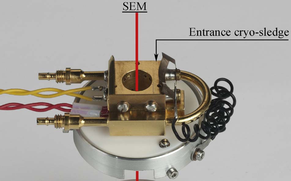
Figure 2456. Sample stage in a cryo-FIB-SEM system. Adapted from [3]
[1] Al-Amoudi, A., Studer, D., Dubochet, J., 2005. Cutting artefacts and cutting process in
vitreous sections for cryo-electron microscopy. J. Struct. Biol. 150 (1), 109–121.
|