=================================================================================
The collimator in an EDS detector in electron microscopes (EMs) is positioned at the front of the detector and constrains the X-rays entering it into parallel paths. The collimator has important roles. As shown in Figure 2537a, the function of the collimator is to reduce the stray x-rays and backscattered electrons that do not originate from the specimen from entering the detector. In other words, the collimator provides a limiting aperture to ensure that only the X-rays from the area on the specimen being excited by the electron beam are detected, and stray X-rays from the microscope chamber are not included in the analysis.
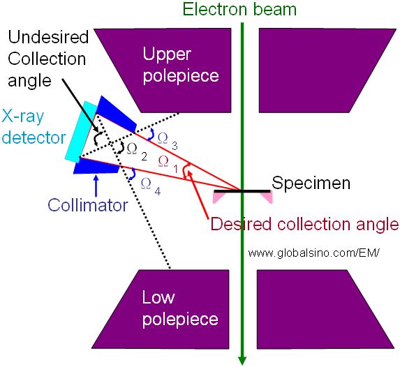
Figure 2537a. Schematic illustration of the correlation between the desired collection angle and collimator in an EDS detector system.
For instance, a design of carbon-coated Pd collimator containing Pb and Al baffles minimizes the transmission of X-rays generated by radiation incident on the outside of the collimator. The Al baffles also constrain the direct entry of backscattered electrons into the EDS detector because the bending of the backscattered electron trajectories by the magnetic field of the objective lens can be used to ensure that the scattered electrons in that path are blocked by the collimator wall or the baffles.
Figure 2537b shows a GIO (grazing incidence X-ray optics) collimator assembly connected to a 10-mm2 EDS detector. The reflectivity of GIOs strongly depends on the grazing angle, material and roughness of reflecting surface, and X-ray energy. The reflecting surfaces are usually composed of a thin, very smooth layer of material such as metals or metal alloys, and have normally different shapes such as cones, parabolas, hyperbolas, ellipsoids, or more complex, combined features. The GIO has the benefits of being compact in size, relatively inexpensive, and highly efficient reflectors for X-ray in the energy range below 1 keV. For instance, the GIO only reflect the X-rays for very small angles of incidence with respect to the reflecting surface. On the other hand, as shown in Figure 2537c, the boron (B), oxygen (O), and carbon (C) peaks have been enhanced by the GIO, comparing with the case without the GIO, while the Nickel (Ni) and silicon (Si) peaks show little or no enhancement.
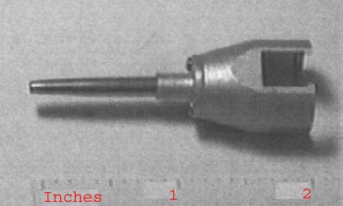
Figure 2537b. A GIO collimator assembly connected to a 10-mm2 EDS detector. Adapted from [1]
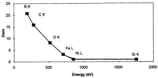
Figure 2537c. The gain enhancement of the GIO collimator as a function of X-ray energy for different elements. Here, the gain represents the ratio of the EDS peaks “with the GIO collimator“ to “without the GIO collimator“. Adapted from [1]
Figure 2537d shows the schematic illustration of an EDS system integrated with a collimator.
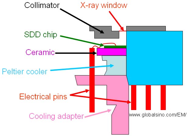
Figure 2537d. Schematic illustration of an EDS system integrated with a collimator.
Note that in EDS measurements, the desired collection angle is set by the collimator placed in front of the detector crystal.
[1] Jon J. McCarthy, and David J. McMillan, Application of X-ray Optics to Energy-Dispersive Spectroscopy, Microsc. Microanal. 4, 632-639, 1999.
|