Chapter/Index: Introduction | A | B | C | D | E | F | G | H | I | J | K | L | M | N | O | P | Q | R | S | T | U | V | W | X | Y | Z | Appendix
|
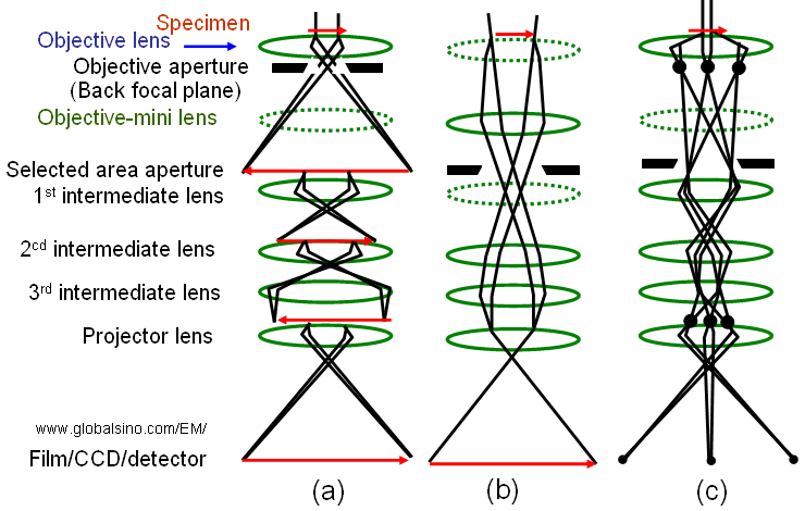
The magnification in TEM can be adjusted in a wide range (e.g. from x50 to x20, 000, 000) by changing the strength of the magnetic field in the magnetic lens and thus changing the focal length. Figure 4140(a) shows the principle of magnifying an image (Normal-Mag mode). A transmitted image of the specimen is first formed and magnified by the objective lens, and then is magnified further by two to four lenses, including an objective lens, intermediate lenses, and a projector lens. As shown in Figure 4140(b), at extremely low magnification (e.g. used for survey of interest), the image is formed by the OM (objective-mini) lens, intermediate lenses, and projector lens. The Diff mode in Figure 4140(c) presents an electron diffraction pattern. In the Normal-Mag mode, the focus of the 1st intermediate lens is adjusted to the image plane of the objective lens where a selected area aperture is located. However, in the Diff mode, the focus of the 1st intermediate lens is adjusted at the back focal plane of the objective lens. Table 4140 lists the status of the lenses and apertures in different operation modes.
Figure 4140. (a) Normal-Mag mode, (b) Low-Mag mode, and (c) Diff mode. The dashed-lenses are turned off in the relevant operation mode. Table 4140. Status of the lenses and apertures in different operation modes*.
In most electron microscopes (EMs), the adjustments of camera length and image magnification use the same knob, that is, different camera lengths are available by turning the magnification knob but this operation does not affect the imaging magnification when you return to image mode. The magnification should be calibrated at the film plane. To achieve a magnification and thus dimension measurement exactly as indicated in the instrument, we need to make sure the operation as follows: Hysteresis factor affects the TEM operations under changed lens conditions once magnetized lenses retain a certain amount of residual magnetism. This means that the level of current used does not always specify lens strength. This effect can particularly affect: i) and ii) can be corrected by pressing the STANDARD FOCUS bottom a number of times during the operation. In this way, the applied current is essentially returned to zero a number of times. This reduces lenses to the standard level of magnetization. In TEM measurements, the magnification of the image-forming lens system has normally an error of 5% - 10% so that the final magnification has an error of 5%-10%. To obtain more accurate magnification (and thus dimension measurement), the magnification should be calibrated with a lattice image of a known standard specimen by recording both the specimen and standard in the same field of view. Furthermore, the instruments may give a few percent of image distortion which also needs to be calibrated. Note that a tip is that focusing the image is often easier at low magnifications than at higher magnifications. Furthermore, the image rotates with the change of magnification because the total magnetic flux strength of the lenses changes. Recently, image rotation-free TEMs have been developed by keeping the total flux strength constant even if the magnification is changed. The magnification and camera length projected on the fluorescent screen are slightly smaller than the ones on the film because the fluorescent screen is located at a higher position than the film.
|