Chapter/Index: Introduction | A | B | C | D | E | F | G | H | I | J | K | L | M | N | O | P | Q | R | S | T | U | V | W | X | Y | Z | Appendix
| One solution to the energy spread (ΔE) of electrons irradiating the specimen in EMs is to use a monochromator in the illumination system. In monochromators, a small magnetic prism and energy selecting slit are installed directly below the source for the purpose of restricting the energy distribution of the primary electron beam. In other words, this setup is essentially an energy filter, consisting of an electron spectrometer operated at a fixed excitation, followed by an energy-selecting slit that restricts the velocity range of the electrons. This strategy is to filter the electron beam close to the electron source, before the incident electrons have been accelerated. Several designs for such gun monochromators have recently been implemented into modern EMs. The aim is to narrow ΔE down to a value which does not affect the measurements in the specific EMs. Assuming the energy spread of the electrons from a monochromator is 0.2 eV, with an accelerating voltage of 200 keV the spectrometer must have an energy resolution and stability of 1 ppm, which require severe stability of the spectrometer and high-voltage power supplies. The quality of EM analysis will be significantly improved if a electron-beam monochromator is used. For instance, EELS typically offers an energy resolution of 1 eV but close to 0.1 eV with an monochromator. Corrections of spherical aberration and off-axial coma have been recently used for improving the spatial resolution of TEM imaging. However, these corrections cannot further improve the resolution when reaching 0.5 Å at voltages ≤ 200 kV due to chromatic aberration. Fortunately, the chromatic aberration can decrease significantly by employing a monochromator or be corrected by crossed electric and magnetic quadrupole elements. In these setups, their electric and magnetic forces compensate each other for the electrons with nominal energies. Up to now, the use of a monochromator and a hexapole aplanator is most promising and holds the chromatic aberration to a minimum and within acceptable limits. Note that there are different types of monochromators currently used in EMs, e.g. Wien-filter type [1]. Furthermore, the energy spreads in the same microscope can be different at different accelerating voltages. For instance, in a microscope with a Schottky-type field emission gun at energy spread of 0.8 eV (full width at half maximum, FWHM) with an accelerating voltage of 300 kV, the electron monochromator reduced the energy spread to 0.13 and 0.08 eV at 300 and 80 kV, respectively. [2] Furthermore, a few monochromators for TEM are now commercially available. Figure 4781 shows the ZLPs (zero-loss peaks) of a CFEG (cold field emission gun) [3] at various emission currents as well as the monochromatized ZLP [4] in logarithmic vertical scale. The inset lists the extraction voltages V1, probe currents Ip and the FWHMs (full width at half maximum) of the ZLPs. Note that the FWHMs quantify the energy spread. In general, the lower extraction voltage and probe current gives smaller FWHMs of the ZLPs, meaning that the higher energy resolution can be obtained, while the one with a monochromator still has the lowest FWHM.
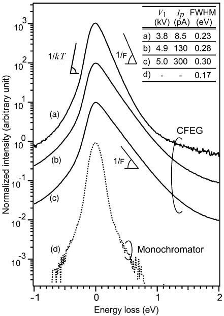
Figure 4781. ZLPs of CFEG under various emission conditions as well as the monochromatized ZLP.
Adapted from [3]
The Fowler-Nordheim (F-N) energy distribution of emitted electrons includes two distinct regions [5]: a low energy Fermi tail and a high energy tunnelling tail. In other words, the energy distribution of emitted electrons is the product of the Fermi–Dirac distribution function ln(1+eΔ/kT) and the tunneling probability exp[-b(φ+Δ)3/2/F], which produce the Fermi tail and the tunneling tail, respectively, and causes beam energy-broadening in field emission guns. The low energy Fermi tail is a property of the Fermi surface of the tip material (e.g. tungesten) and is independent of the extraction voltage. The left side of the CFEG’s ZLP is comparable to that of the monochromator. The slope of the high energy tunnelling tail is determined by the field strength. In this case, it is difficult to determine the onset of the inelastic scattering within a low-loss spectrum because the tunneling tail induces a strong background. Therefore, the monochromator has an advantage in the measurement of small bandgaps (e.g. ≤1 eV).
[1] Tiemeijer, P.C. (1999). Measurement of Coulomb interactions in
an electron beam monochromator. Ultramicroscopy 78, 53–62.
|