Chapter/Index: Introduction | A | B | C | D | E | F | G | H | I | J | K | L | M | N | O | P | Q | R | S | T | U | V | W | X | Y | Z | Appendix
| Among charged particles, electrons possess the smallest mass, which minimizes the structural damage that they cause in the specimen. However, when energetic electrons interact with SEM samples or pass through a thin film in TEM, they still lose their energy, mainly through heating, electrostatic charging, displacement damage, sputtering, ionization damage (radiolytic), inelastic collision (knock-on), and hydrocarbon contamination processes [1], as shown in Figure 4543a. These phenomena are often called “radiation damages” in a less precise form. In EM analysis, ionization damage and sputtering are more common than displacement damage. Here, the radiation damage refers to any changes in physical structure and chemical composition which occur as a result of exposure to the electron beam. The extent of electron-beam damage in electron microscopes, e.g. high voltage TEMs, depends on some factors such as the probe current density, accelerating voltage, chemistry and structure of the solid, and type and concentration of defects in the specimens. Almost all materials undergo the displacement of atoms above a specific energy threshold, and electrons with energies smaller than this critical displacement energy only make the sample atom vibrate in its site and dissipate energy as phonons. Direct displacement of atoms from the crystal lattice creates point defects. In radiolytic damages based on the inelastic scattering, an increase in excitation energy can produce bond breaking of certain materials such as polymers and alkali halides. The damages which affect the structure and/or the chemistry of the specimen depend mainly on the energy of the electron beams. The ionization effect decreases significantly with increasing accelerating voltage up to 100 kV and stays low at higher voltage [2]. Therefore, when a material can undergo a radiolytic process, knock-on damage is insignificant and vice versa. For most metals, for example, the threshold of displacement energy is about 20 to 30 eV [1]; therefore knock-on damage in the TEM does not occur for accelerating voltages less than 300 kV.
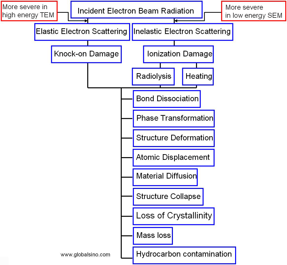
Figure 4543a. Schematic diagram of radiation damage. In TEM analysis, most ceramics and catalysts suffer radiation damages under electron beams with accelerating voltages of 80-300 kV (especially in FEG TEMs due to high current densities), while most of metallic materials are not affected by such incident electron beams. Figure 4543b shows the details of the different types of electron beam radiation damage. The direct atomic displacement, so-called knock-on damage, which is generated by the high energy electrons due to large momentum transfering, normally occurs only during TEM analysis due to the high acceleration voltages of up to 400 keV. Knock-on damage can happen in organic compounds but is not the major contributor to radiation damage of organics due to the faster and significant ionization damage before such knock-on damage could occur. Table 4543. Degree of radiation damage of different materials.
Figure 4543b. Detailed classification of electron beam radiation damage. Note that radiation damage from the electron beam is a complex phenomenon and thus it is impossible to determine the details about the damage in real-time while data collection is in process.
[1] L. W. Hobbs, in:J. J. Hren, J. I. Goldstein, D. C. Joy (Eds.),Introduction to Analytical Electron Microscopy, PlenumPress, NewYork, 1979, p.437.
|
|||||||||||||||||||||||||